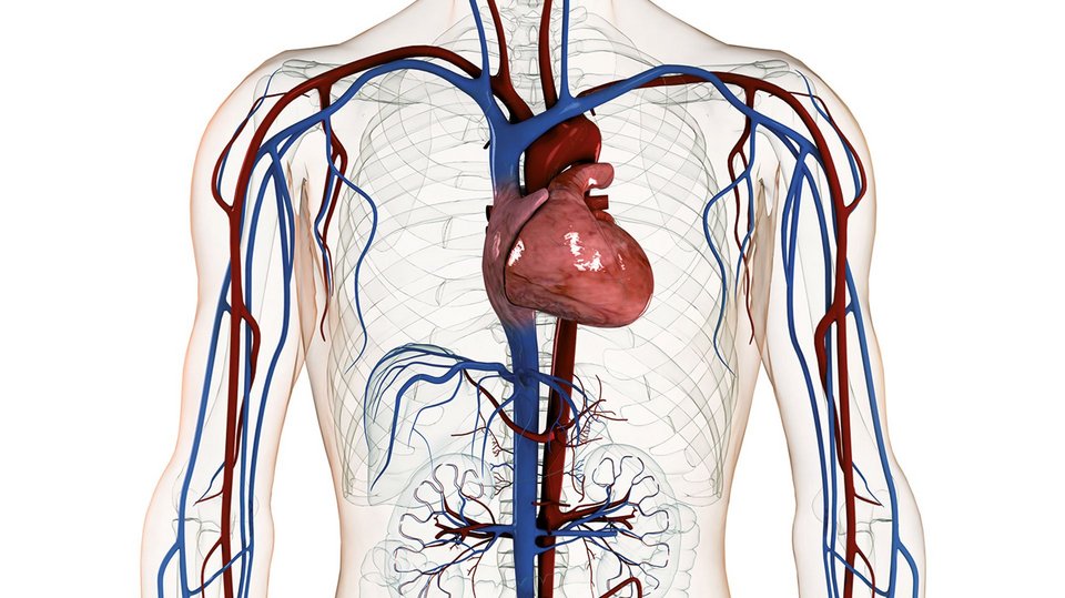
Cardiology deals with disorders of the heart (conducting system, coronary vessels, heart muscle and heart valves).
The field includes both non-interventional and catheter-assisted interventional procedures. The modern equipment of the Cardiovascular Center Klinikverbund Allgäu together with the first hybrid catheter laboratory (catheter laboratory and operation theatre in one room) in the southwest of Germany makes the outpatient and inpatient examination and treatment of patients by means of the most innovative, interventional procedures possible.
Together with the Doppler echocardiography, echocardiography is the most important non-invasive imaging technique in cardiology. It is a means of diagnosis of morphologic changes of the heart and it provides functional information in case of cardiac diseases such as atrium, ventricle and valve disorders. The transoesophageal echocardiography (TEE) provides additional information on morphologic changes of the valves (e.g. bacterial appositions = vegetations), lacerations of the ascending thoracic aorta and on the existence of blood clots especially in the left atrium and left atrial appendage.
The resting ECG is an indispensable tool for the diagnosis of the coronary heart disease, cardiomyopathies (heart muscle diseases), valve disorders and especially cardiac arrhythmias. The exercise ECG serves to measure the resilience of patients and the response of the heart circulation system (pulse rate, blood pressure) to the exercise. A long-term ECG records the heart rhythm and possible irregularities during the time of registration. Cardiac asystoly (pause of the heart beat) or bouts of tachycardia (rapid heart beat), atrial or ventricular extra beats and, above all, life-threatening ventricular tachycardias will be detected by these long term holter recordings. The long-term blood pressure measurement is necessary to determine the type and the severity of the blood pressure disorder and to check medical therapies intended to lower the blood pressure.
The pacemaker and ICD control is meant to check the function of these devices and their electrodes. In addition the functional state of the battery is tested. The programming of the pacemaker is adjusted to the individual needs of the patient and if necessary, adjustments are made.
In ICD patients the defibrillator function will be checked in addition (i.e. interrogation of the tachycardia and ICD- therapy storage), and readjustments of the device will be performed if necessary.
Medicinal therapies
- the conservative, medicinal treatment of the coronary heart disease can prevent the progress of the disease and it can reduce its impact on the heart
- conservative, medicinal treatment of the ischaemic and non-ischaemic heart insufficiency as well as cardiac arrhythmia
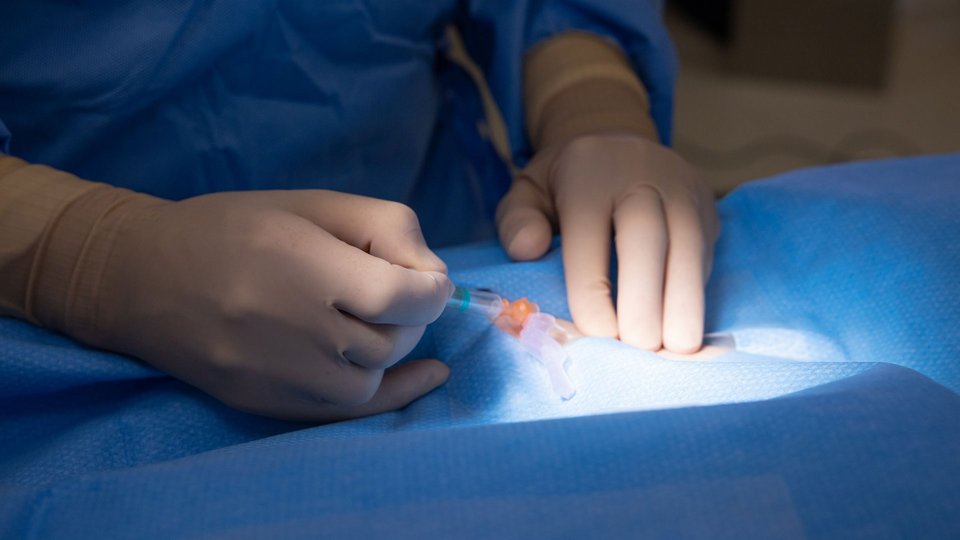
During the examination of a heart catheter, the heart and the coronary vessels are examined by means of the X-ray method. In this procedure, a tiny plastic tube is inserted into an arm or groin area artery and guided to the left ventricle. Furthermore, a contrast agent is administered to make the vessels become visible on the X-ray monitor.
By doing so, the coronary vessels and the activity of the valves can be visualized. Thus, the heart specialist can spot possible constrictions and repair them if necessary.
The Percutaneous Transluminal Coronary Angioplasty (PTCA) is a therapeutic procedure used to expand narrowed coronary arteries by placing a balloon catheter at the constriction in the vessel. A balloon catheter is a thin, flexible plastic tube with an inflatable balloon at its end. There is no major surgery needed for the implantation of the vessel supports (stents). A cut into a groin or wrist artery is sufficient. From there, a fine wire is used to push forward the stent to the constriction expanded by the balloon in the blood vessel. The stent helps preventing the artery from narrowing again.
In the event of what are termed intermediate stenoses, that is, borderline significant narrowing of the coronary blood vessels for the supply of blood to the heart muscle, FFR measurement in the course of heart catheter investigation is the gold standard for ischemia diagnostics (assessment of the corresponding relevance of the stenosis). This involves using a pressure wire to measure a pressure differential between the vascular section in front of and behind the stenosis at maximum dilation of the blood vessel. If a haemodynamically relevant stenosis is apparent, this can be immediately treated via the pressure wire with a balloon dilatation and/or stent implantation.
This is a catheter based procedure during which a new aortic valve is transported through the groin to its intended position in the heart. The biological valve (porcine or bovine) is placed on a stent which is unfolded when expanding the narrowed aortic valve. Then, the new valve is in the stent on its original position.
A new approach to further examine unclear disorders of the cardiac function is the so-called myocardial biopsy. During this procedure, small samples are taken out of the heart muscle at the top of the right ventricle. These samples enable differential histological and molecular-biological examinations. The diagnosis of a chronic virus infection of the heart is of special importance because then, antiviral treatments with Beta-Interferon 1B for example can be taken into consideration.
The pacemaker can repair various disorders in the electric conduction system of the heart by sending an electric impulse in case of deceleration of the heart rhythm or temporary cardiac arrest. The impulse causes the heart muscle to resume regular contractions. A defibrillator is some kind of pacemaker which is able to terminate ventricular tachycardia as well as ventricular fibrillation. This is done by sending a quick impulse to the ventricles or, if necessary, by using electroshocks.
A residual slit-like opening between the atria (PFO = patent foramen ovale) can be a possible cause of a stroke. If permanent thinning of the blood required for this reason (oral anti-coagulation e.g. with Marcumar) is not possible or wanted, the PFO can be closed with a kind of small umbrella (PFO occluder) in the heart catheter laboratory. This process can also be performed to occlude an atrial septal defect (ASD) which, if untreated, can result in right-sided heart failure with corresponding shortness of breath at rest and during exertion.
The Cardiovascular Center Klinikverbund Allgäu carries out inpatient heart catheter examinations, too. A conversation with the patient will show whether an inpatient heart catheterization is appropriate or not. This might be necessary in case of concomitant diseases such as kidney or lung diseases and when there is a high risk of complications during the heart catheterization.


![[Bitte in "english" übersetzen:] [Bitte in "english" übersetzen:]](/fileadmin/_processed_/3/6/csm_Herz-Gefaesszentrum_Kliniken_b881e41a9d.jpg)
